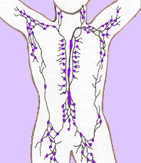
lymphoid system
 Components of the lymphoid system are:
● immune cells – B cell lymphocytes and plasma cells, dendritic cells, granulocytes, macrophages, monocytes of mononuclear phagocyte system (MPS), T cell lymphocytes
● lymph ducts and vessels and lymph nodes (right)
[] histopathology []
● lymphoid organs including reticuloendothelial system – bone marrow, (lymph nodes), mucosa-associated lymphoid tissue, Peyer's patches, spleen, thymus, tonsils, vermiform appendix
The reticuloendothelial system or mononuclear phagocytic system comprises a range of cells that are capable of phagocytosis, including macrophages and monocytes. Phagocytosis is an innate rather than an adaptive immune process. The phagocytic cells either circulate in the blood or are attached to various connective tissues such as pulmonary alveoli, liver sinusoids, skin, spleen, and joints.
The RES functions to provide phagocytic cells for both the inflammatory response and immune responses (primary RES) and to remove pathogens and senescent cells from circulation (secondary RES)
The reticuloendothelial system (RES) includes:
● primary (central) lymphoid production organs – bone marrow, thymus
● secondary (peripheral) lymphoid function organs – circulating monocytes, histiocytes located in many tissues, Kupffer cells of the liver, "Littoral cells" of the spleen, mucosa-associated lymphoid tissue (MALT), which is subdivided into bronchus-associated lymphoid tissue (BALT) and gut-associated lymphoid tissue (GALT), Peyer's patches.
o-o index of tissue micrographs [] germinal centers [] fluorescence microscopy dendritic cell uptake of dying cells within spleen [] micrograph red pulp of spleen [] micrograph splenic red pulp [] micrograph spleen cells (mouse) DAPK2 stained [] micrograph white pulp, splenic nodule [] micrograph white pulp infiltrate with Langhans giant cell [] histopathology spleen Gaucher's disease [] micrograph gallery cell surface antigens [] Virtual Histology Main []
▲ Top ▲
tags [Tissue] [lymphoid] [reticuloendothelial] [lymph nodes] [thymus]


